
A foundation model for IHC biomarker scoring: one pass, every cell found and typed, across PD-L1, ER, PR, HER2 and Ki-67.
Immunohistochemistry is the workhorse of precision oncology. A single brown stain decides whether a patient is offered immunotherapy, endocrine therapy or an anti-HER2 drug. Yet behind every PD-L1 TPS, every Ki-67 index and every HER2 category sits the same slow step: someone has to look at thousands of cells and decide which ones count.
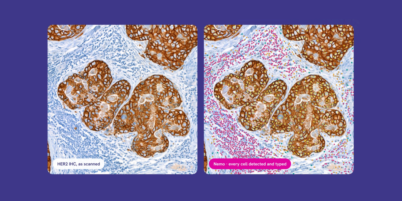
Today we are introducing NEMO, our foundation model for cell-level IHC analysis. NEMO detects every cell on an IHC slide, types it, and calls its marker status, using one model across five markers with no retraining between them.
Why IHC Scoring Is Harder Than It Looks
IHC scoring looks objective: a cell is brown or it is not. In practice it is anything but. Staining intensity varies between labs, antibody clones and scanners. Membranous, cytoplasmic and nuclear patterns overlap. And pathologists scoring the same slide routinely land on different sides of a clinical cut-off, especially near the low thresholds that matter most, like PD-L1 TPS at 1%.
Most AI tools for IHC inherit this problem directly. They are trained on what annotators clicked, so they learn the annotators' habits along with the biology. As we wrote in About Ground Truth in AI Single-Cell Detection, a model graded against the same labels it learned from can look excellent on paper and still drift in the real world.
The usual answer is one model per marker, each trained on its own hand-annotated dataset. That approach is slow to build, expensive to maintain, and caps every model at the agreement level of its annotators. We wanted to build something different.
Built on Molecular Ground Truth
Instead of asking humans what each cell is, NEMO learns from what each cell expresses. We pair routine IHC slides with a direct molecular readout taken from the same tissue section, then let that measurement decide the label.
The pipeline has four steps:
- Stain. Start from a routine IHC slide: any marker, any assay.
- Measure. Acquire molecular ground truth on the same section, cell by cell.
- Resolve. Turn each cell's marker signature into a cell state: tumour positive, tumour negative, immune positive, immune negative, or stroma.
- Transfer. Map those states back onto the nuclei in the IHC image.
The result is an IHC database of 2.3 million molecularly labelled cells across five markers, and it is what NEMO is trained on. Because the labels come from the tissue's own biology rather than a reader's judgement, the same recipe applies to any marker we add, without starting a new annotation campaign each time.
How NEMO Performs, Marker by Marker
We evaluated NEMO on all five markers against human references. Cell detection is validated on every marker; for PD-L1, scoring is validated too.
| Marker | Evaluation set | Reference | Detection F1 | Precision | Recall | Scoring |
|---|---|---|---|---|---|---|
| PD-L1 | IGNITE: 32 regions, 9,473 nuclei | 4 pathologists | 0.81 | 83.4% | 79.1% | Validated |
| ER | EndoNuke: 148,210 nuclei | 7 annotators | 0.876 | 91.5% | 84.0% | In evaluation |
| PR | EndoNuke: 58,852 nuclei | 7 annotators | 0.869 | 90.9% | 83.2% | In evaluation |
| HER2 | Tumour region | 1 pathologist | 0.984 | 96.8% | 100% | In evaluation |
| Ki-67 | Tumour region | 1 pathologist | 0.934 | 98.7% | 88.7% | In evaluation |
PD-L1: detection and scoring validated
PD-L1 was evaluated on IGNITE, a public multi-reader dataset spanning two assays and two centres, with four pathologists as reference. Evaluation is at region level, not whole slide.
Detection. Across 32 regions and 9,473 nuclei, NEMO reached an F1 of 0.81, ranging from 0.80 to 0.82 depending on which pathologist it is scored against. For context, the pathologists agree with each other at 0.85 to 0.88.
Per-cell status. Across 98 regions and 5,650 tumour cells, NEMO called PD-L1 status with 89.7% balanced accuracy (ROC AUC 0.96), with 87.0% sensitivity and 92.4% specificity.
Tumour proportion score. On 23 regions scored for TPS, NEMO's score correlated with the pathologists at r = 0.93, with a mean absolute error of 9.7 points and an average bias of just +0.8 points; the 95% limits of agreement span ±33.3 points, so individual regions can still differ substantially. Agreement at the clinical cut-offs was 78.3% at TPS ≥ 1%, 95.7% at ≥ 5%, 87.0% at ≥ 10% and 91.3% at ≥ 50%.
ER and PR: within 0.02 of human agreement, by median F1
On EndoNuke, annotated by three pathologists and four medical students, NEMO reached an F1 of 0.874 across 210,419 nuclei: 0.876 on ER (148,210 nuclei) and 0.869 on PR (58,852 nuclei), with the remaining 3,357 nuclei coming from tiles whose stain was not labelled (F1 0.890). In a subset of 20 tiles labelled by all seven annotators, NEMO agreed with each annotator at a median F1 of 0.89. The annotators agree with each other at a median of 0.91.
HER2 and Ki-67: near-complete detection
In the tumour region, NEMO found every epithelial cell the pathologist annotated on HER2 (recall 100%, F1 0.984). On Ki-67, precision reached 98.7% with an F1 of 0.934. HER2 category scoring and Ki-67 index scoring are now being evaluated with our clinical collaborators, as is ER/PR positivity scoring.
One Model, Every Marker
NEMO is designed around three ideas.
Assay-agnostic by design. NEMO handles PD-L1, ER, PR, HER2 and Ki-67 zero-shot, with no retraining between them. Membranous, cytoplasmic and nuclear staining patterns are all handled by the same model.
Every cell, not a sample. NEMO scores every detected cell in the region, not a hand-picked field. Tumour, immune and stromal cells are typed in the same pass, so a score is always traceable to the cells behind it.
Grounded in biology. Because NEMO learns from molecular measurements rather than visual consensus, its notion of "positive" is anchored to what cells actually express. That is what lets a single model generalise across markers and assays.
For pharma and CRO partners, this means one consistent scoring engine across trials and biomarkers. For laboratories, it means a quantitative, reproducible second read on the slides they already produce.
What's Next?
We are now completing scoring validation for ER/PR positivity, HER2 category and Ki-67 index with our clinical collaborators, and extending NEMO to further markers.
Send us your toughest slide. We will show you what NEMO makes of it.