

Improved cell classification across every evaluated cell type, with a new lightweight model for the research community.
At Octopath AI, we believe the future of computational pathology lies in understanding tissue at the cellular level. Every tumour contains a complex ecosystem of cancer cells, immune cells, stromal cells, and vascular structures. Understanding their composition, spatial organisation, and interactions is essential for advancing cancer research, biomarker discovery, and personalised treatment.
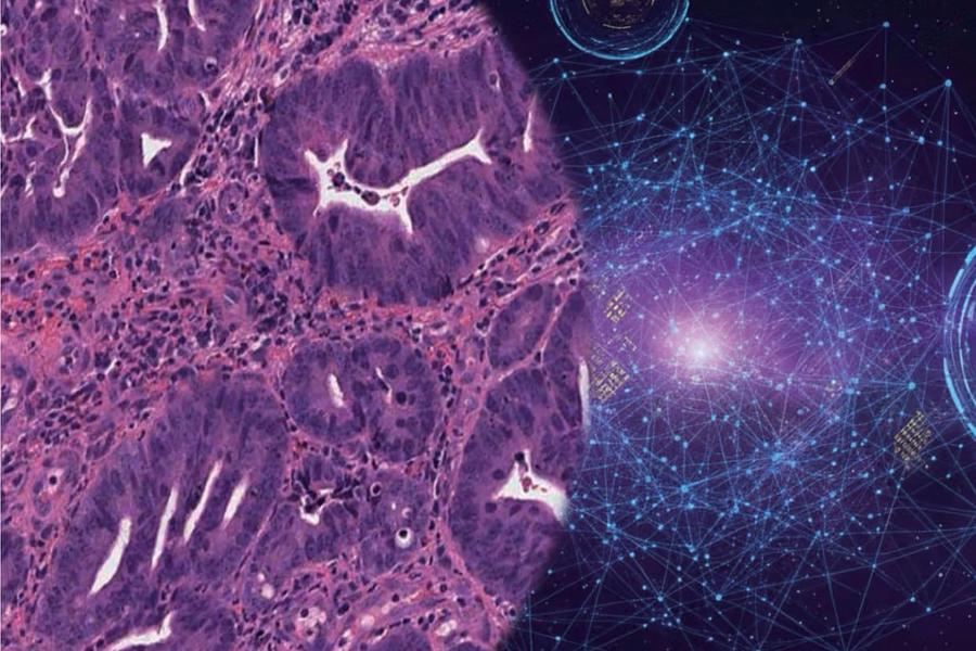
This is the vision behind Kraken-1, our AI foundation model for cell-level analysis of routine histopathology images.
With our October 2026 update, Kraken-1 takes another step forward, delivering improved classification accuracy across all eight evaluated cell types. We are also introducing a compact version of the model, Kraken-Open, to make our technology more accessible to the research community.
A Foundation Built on 14.1 Million Cells
Earlier this year, we introduced Kraken-1, our AI foundation model designed to detect and classify individual cells directly from routine haematoxylin and eosin (H&E) histopathology images.
Kraken-1 was developed using a large-scale database of 14.1 million labelled cells spanning 14 tissue types, including colorectal, breast, lung, brain, and liver tissues. By learning from diverse tissue environments, the model aims to capture shared cellular features while accounting for morphological differences across organs and cancer types.
Since its introduction, we have continued refining Kraken-1, with a particular focus on improving classification accuracy for challenging cell populations.
What's New in October 2026?
Our latest model update delivers consistent improvements across all eight evaluated cell types, increasing the overall macro F1 score from 55.7% to 63.8%, an improvement of 8.1 percentage points, on our validation dataset.
The most substantial gains were achieved in cell populations that are particularly difficult to distinguish based on morphology alone:
- Vascular cells: +16.6 percentage points
- Smooth muscle cells: +14.0 percentage points
- Neutrophils: +7.5 percentage points
- Macrophages: +7.4 percentage points
We also achieved improvements in epithelial cells, lymphocytes, fibroblasts, and plasma cells. The updated model reaches an F1 score of 88.4% for epithelial cells and 81.0% for lymphocytes.
These results reflect meaningful progress in distinguishing morphologically similar cell populations, particularly within the stromal and vascular compartments, which remain challenging for automated histopathology analysis.
Towards More Comprehensive Tissue Understanding
Accurate cell identification is only the first step. Our broader goal is to transform routine histopathology images into quantitative maps of tissue biology.
Kraken-1 is designed around three key capabilities:
Accurate cellular identification. Detecting and distinguishing epithelial, immune, stromal, and vascular cell populations provides a foundation for detailed tissue characterisation. The latest improvements are particularly encouraging for cell types with overlapping morphological features.
Cross-tissue applicability. By learning from millions of cells across multiple organs, Kraken-1 aims to provide a unified approach to cellular analysis across different tissue and cancer types, reducing the dependence on tissue-specific models.
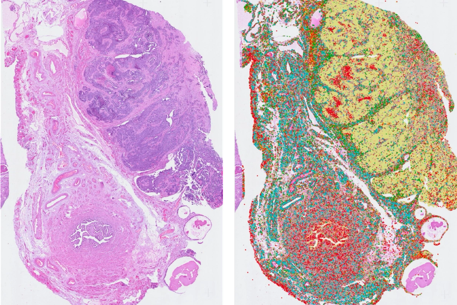
Scalable quantitative pathology. Kraken-1 enables cell detection, classification, and spatial mapping directly from routine H&E slides, supporting applications such as tumour cellularity assessment, immune microenvironment profiling, tumour–stroma analysis, and quantitative evaluation of tissue architecture.
Together, these capabilities bring us closer to extracting richer biological information from the histopathology slides already generated in routine clinical practice.
From Cell Detection to Precision Oncology
Our ambition extends beyond identifying individual cells.
The tumour microenvironment plays an important role in cancer progression and treatment response. By quantifying the abundance, distribution, and spatial relationships of different cell populations, Kraken-1 provides a foundation for investigating biomarkers associated with patient prognosis and therapeutic outcomes.
This is particularly valuable in clinical research, where H&E slides are widely available, while comprehensive molecular profiling may be constrained by cost, tissue availability, and technical complexity.
We envision Kraken-1 supporting large-scale studies of tumour biology, helping researchers uncover cellular and spatial patterns that could inform patient stratification and personalised treatment strategies.
Introducing Kraken-Open: A Taste of What's Possible
We are excited to introduce Kraken-Open, a compact version of Kraken-1 designed for non-commercial research use. By making advanced cell-level pathology AI more accessible, we aim to empower the research community to explore new applications, independently evaluate model performance, and accelerate innovation in computational pathology.
Explore Kraken-Open on Hugging Face:
Interested in unlocking the full potential of single-cell tumour microenvironment analysis with Kraken-1? Please contact us to explore access to the full model and discuss how we can support you.
What's Next?
Our work on Kraken-1 is continuing. Alongside further model development, the research paper detailing Kraken-1's development and performance evaluation is coming soon.
We are excited about the progress made so far, and this is only the beginning.
Stay tuned for our upcoming research paper and further model updates.