
Octo Mitosis
Mitotic figure detection and hotspot mapping on H&E
Mitotic figure detection and hotspot mapping from routine H&E, reported per 10 HPF and per mm² with the cells it counted.
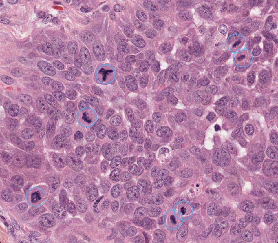
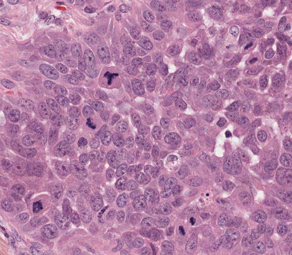
Manual mitotic counting is slow and variable.
Finding the hotspot and counting mitoses in ten high-power fields takes minutes per slide and varies between pathologists and between days. Under-counting shifts a grade; over-counting shifts a treatment.
The count feeds tumour grade. Mitotic activity is a component of grade in breast carcinoma, sarcoma and neuroendocrine tumours, and of proliferation indices across oncology. A reproducible count is a reproducible grade.
Three things you stop doing by hand
- 01Every figure found
86% pan-cancer mitosis detection accuracy in the Kraken benchmark, learned from 14.1 million labelled cells; atypical figures flagged separately.
- 02A count that does not drift
The same count for the same slide, every reader, every day. Hotspot chosen by the model, shown for you to confirm.
- 03The true hotspot
Whole-slide search, not ten fields: the hotspot is found across the entire tumour, not where the eye happened to land.
The densest 2 mm², found for you
Octo Mitosis reads every field on the slide, not ten chosen by eye, then locks onto the area with the most mitotic figures and reports the count per 2 mm².
Used for grading in breast cancer, GIST, meningioma, neuroendocrine tumours and sarcoma.
What you get back
Output | What you get | Format |
|---|---|---|
| Mitotic count | per 10 HPF and per mm², with the field diameter used | data |
| Hotspot map | the densest 2 mm² region, outlined on the slide | overlay |
| Atypical mitoses | flagged and counted separately | data |
| Per-cell export | coordinates and class for every detected figure | CSV / JSON |
| Report snippet | count, basis and hotspot thumbnail for the pathology report |
Mitotic count per 2 mm², as in the WHO Classification of Tumours and RCPath reporting standards · Reference →
Evidence
86% is the Kraken pan-cancer benchmark (UCL, 2026). Regulatory status: research use only. Reference protocol: 10 HPF (2 mm²) in the hotspot.
Count mitoses against the epithelium, not the area
Pancreatic cancer is a ductal disease, so Octo Mitosis also reports mitotic figures per 1,000 epithelial cells, using the epithelial cells Kraken finds. On the same slides, that ratio carries more prognostic information than the conventional 10-HPF count.
Filled square: 95% CI excludes 1. Cox models adjusted for age and stage, one diagnostic slide per patient, stage IV excluded for PDAC. OS overall, DSS disease-specific, DFS/PFS disease- or progression-free survival.
Adding the epithelial ratio to the 10-HPF count improves the disease-free survival model on the same patients (likelihood-ratio test):
- TCGA-PAAD · DFSχ² 5.7, p = 0.017
The same ratio over the whole tumour is prognostic for overall survival in breast (TCGA-BRCA, HR 1.25) and lung adenocarcinoma (TCGA-LUAD, HR 1.18), adjusted for stage and, in breast, PAM50 subtype.
Not replicated in CPTAC-PAAD (unadjusted, p = 0.64–0.96); robustness across cohorts and pipelines is being studied.
Try Octo Mitosis
Run it on a slide in the platform, or ask for the product sheet with the full output specification.