
Decoding Cancer: How AI Counts Cells at Lightning Speed
The Challenge
Tumour cell density, in the tumour region, is a key marker that helps us predict the tumour aggressiveness and its response to a variety of treatment agent.. Pathologists have traditionally done the painstaking work of estimating this density, spending 20 minutes per slide, manually estimating cells in selected 9 mm² regions. It's time-consuming, subjective, and frankly, not scalable for the hundreds of patients seen per a Trust per year. We can do better with AI.
A Two-Stage Detection System
Our proposition lies in a clever dual-AI framework that mimics human pathology assessment, a tag-team approach to cell detection.
Stage 1: The Tissue Classifier
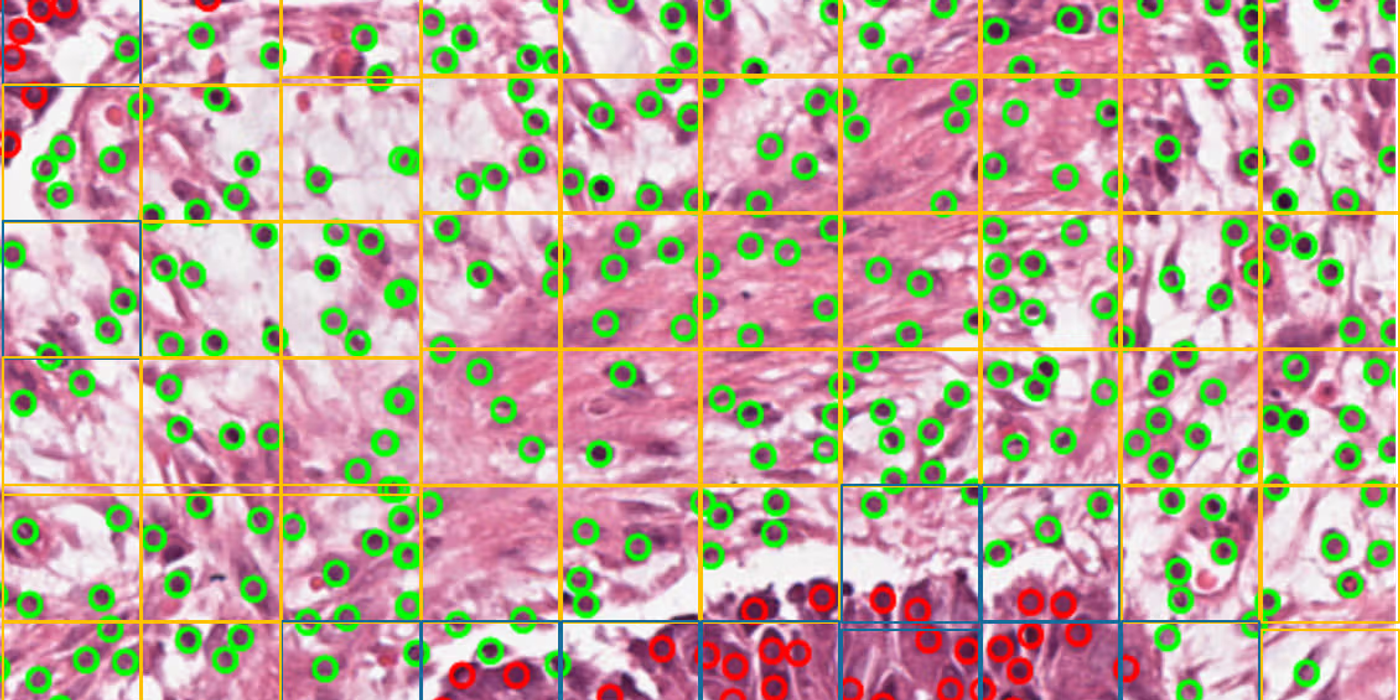
The first AI is an EfficientNet neural network, trained on the NCT-CRC-HE-100K dataset—a massive collection of 100,000 colorectal tissue images. This classifier acts as a sophisticated tissue cartographer, segmenting whole-slide images into nine distinct tissue types, including the critical duo: tumour and stroma.
Stage 2: The Cell Detective
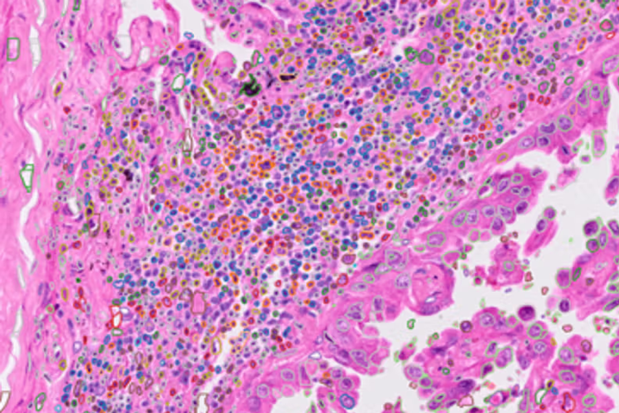
Our second AI model detects and classifies individual cells as either tumour or non-tumour. This cell-level precision is what sets this approach apart from cruder tile-based methods.
Speed and Scale
The performance metrics are impressive:
- <1 minutes per whole-slide image
- Near real-time analysis compared to 20 minutes manually
- Processes entire slides, not just selected regions
And above and beyond, we have demonstrated a very high Concordance with manual assessment: CCC = 0.831 (95% CI: 0.760–0.886).
Why Cell-Level Detection Matters
Previous AI approaches classified tissue tiles—essentially dividing the image into squares and labeling each square as "tumour" or "stroma." The problem? Tiles contain mixtures. A "tumour tile" might have stromal cells, and vice versa, limiting precision.
By detecting individual cells within already-classified tissue regions, this framework achieves unprecedented granularity. You're not estimating—you're counting.
Validation That Counts
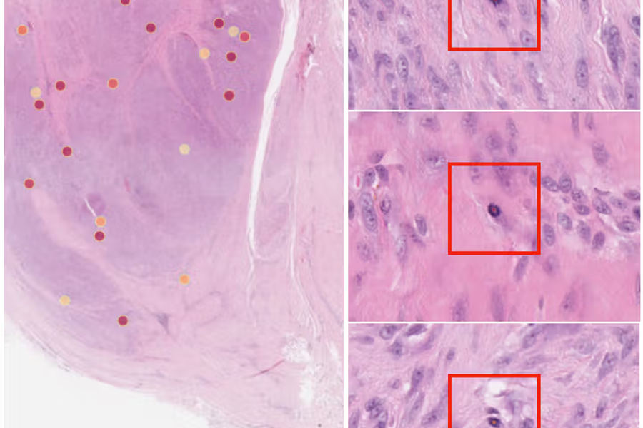
The framework was validated against 116 manually annotated whole-slide images. The Bland-Altman analysis revealed a mean bias of just -0.048, with tight 95% limits of agreement (-0.254 to 0.158). In plain English: the AI and pathologists are seeing nearly the same thing.
The Clinical Promise
This isn't just a technical achievement—it's a clinical tool ready for deployment. The framework is:
- Compatible with existing digital pathology systems
- Scalable to large clinical trials
- Reproducible, eliminating inter-observer variability
- Open source (available on GitHub)
What took pathologists 20 minutes now takes an algorithm 2 minutes, with consistent, quantitative results that can stratify patients for personalized treatment strategies.
The future of precision oncology is not only in discovering new methods, but in making sure that we can see what pathologists see, faster and more consistently than ever before.