
Detecting Cancer's Cellular Signatures: How AI is Revolutionizing Mitotic Figure Detection
Counting dividing cells under a microscope might sound straightforward, but for pathologists diagnosing cancer, it's a painstaking task that can make the difference between accurate treatment and misdiagnosis. Enter OMG-Net (Optimised Mitoses Generator Network) - a breakthrough AI framework that's changing the game.
The Challenge: Finding Needles in Microscopic Haystacks
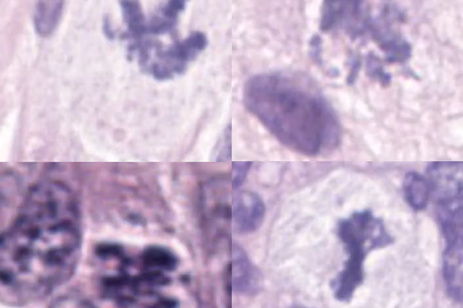
When cells divide during mitosis, they're markers of how aggressively a tumor is growing. Pathologists traditionally count these "mitotic figures" in tissue samples to grade cancers - but there's a problem. These cells are tiny, they look different at various stages of division, and other cellular structures love to masquerade as mitotic figures. Even experienced pathologists often disagree on what they're seeing.
The result? Time-consuming analysis prone to human error, with potential consequences for patient care.
The Breakthrough: A Two-Stage AI Detective
Researchers at University College London and collaborators have created something remarkable: the largest dataset of mitotic figures ever assembled (74,620 examples!) and an AI framework that outperforms previous state-of-the-art models.
Here's what makes OMG-Net special:
1. Segment Anything Model (SAM) as a Cell Finder
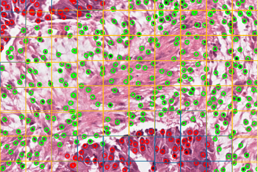
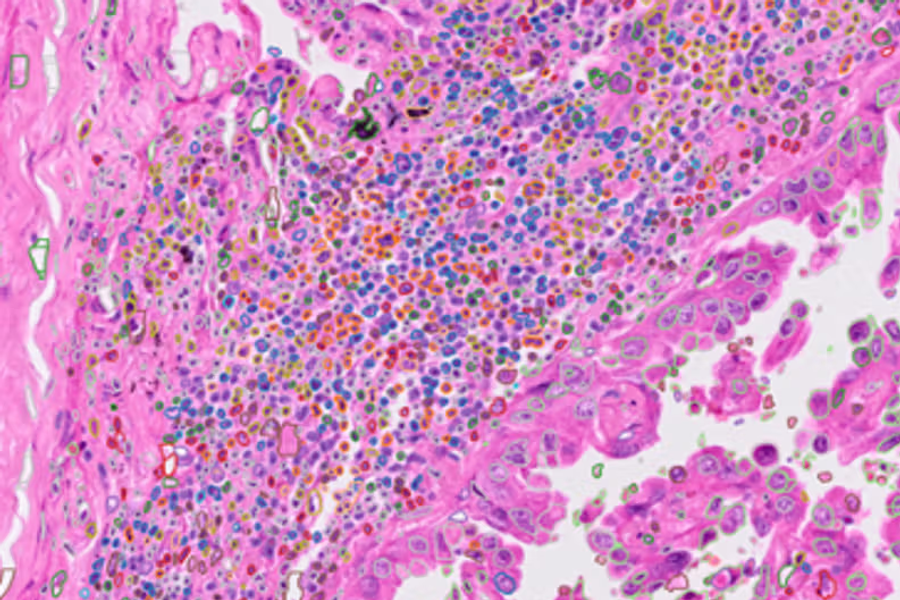
First, SAM automatically outlines every cell-like object in microscope images. Think of it as casting a wide net to catch everything that might be interesting.
2. Smart Classification with Enhanced Vision
Then, an adapted ResNet18 neural network examines each outlined object, but with a twist - it sees both the regular microscope image and the cell outline simultaneously. This dual perspective helps it focus on the morphological features that matter.
The Data Innovation: Building a Better Training Ground
The team didn't just improve algorithms - they revolutionized data collection:
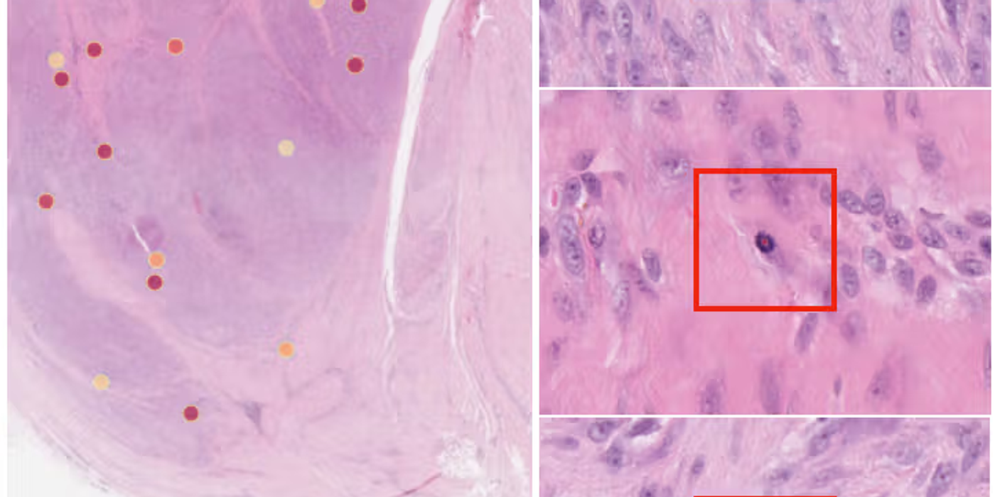
- Smart antibody staining: They used immunohistochemistry (pHH3 antibody) to automatically highlight mitotic figures before manual review
- Active learning loops: Pathologists corrected AI predictions, which fed back to improve the model
- Multi-species insight: Surprisingly, including canine cancer data helped train better models for human cancer detection
The Results: Beating the Competition
OMG-Net achieved an F1 score of 0.84 across multiple cancer types, significantly outperforming existing models on breast carcinoma, neuroendocrine tumors, and melanoma. In head-to-head comparisons with top-performing models from the MIDOG 2022 challenge, OMG-Net consistently came out on top.
Why This Matters
This isn't just about impressive metrics - it's about:
- Consistency: AI doesn't have "bad days" or observer variability
- Speed: What takes pathologists hours can happen in minutes
- Rare cancers: The framework includes the first large dataset for soft tissue tumors, a group of over 100 rare cancer subtypes
- Generalizability: Trained on data from multiple scanners, labs, and tumor types
The Road Ahead
The researchers are refreshingly honest about remaining challenges: neuroendocrine tumors still prove tricky, rare cancer datasets need expansion, and clinical implementation requires careful validation alongside pathologists.
But the foundation is solid. With continuous learning, federated training across institutions, and pathologist-in-the-loop refinement, OMG-Net represents a significant step toward computer-aided cancer diagnosis that could genuinely improve patient outcomes.
The Bottom Line: By combining foundation models like SAM with smart data curation and innovative two-stage detection, OMG-Net shows that AI can handle one of pathology's most challenging tasks - finding and counting the tiny cellular signatures that reveal cancer's true nature.
The code and dataset are open-source and available on GitHub and Zenodo, ensuring the research community can build on this work.