
CytoProfile: Bringing Advanced Cell Analysis to Everyday Pathology Slides
If you've ever wondered why cutting-edge spatial biology techniques haven't made it into routine cancer diagnosis, the answer is simple: cost and complexity. Technologies like multiplex immunofluorescence can reveal incredible cellular detail, but they're expensive, time-consuming, and require specialized equipment. Meanwhile, pathologists worldwide rely on humble H&E (Hematoxylin and Eosin) stained slides - the century-old workhorse of tissue analysis.
What if you could get the best of both worlds?
The Vision: Spatial Biology Meets Routine Pathology
CytoProfile is a deep-learning model with an ambitious goal: to automatically identify and classify different cell types in standard H&E slides, bringing multiplex immuno-fluorescence-level insights to the slides that are already being made in every pathology lab, every day.
Think of it as teaching an AI to see what expensive molecular techniques can see, but using only the basic stains that have been around since the 1800s.
Why This Matters for Cancer Research
Understanding the tumor microenvironment (TME) - the complex neighborhood of cancer cells, immune cells, blood vessels, and supporting tissue - is crucial for:
- Predicting treatment response: Different cell populations can indicate whether immunotherapy might work
- Understanding cancer progression: How cells are organized spatially tells us about tumor aggressiveness
- Personalized medicine: Detailed cellular composition helps tailor treatments to individual patients
Traditionally, getting this level of detail required either:
- Manual annotation by expert pathologists (slow and subjective)
- Expensive multiplex immunofluorescence (not practical for routine use)
How CytoProfile Works
The clever part is the training strategy. CytoProfile learns from high-resolution multiplex immunofluorescence data - which definitively identifies what each cell is - and then transfers that knowledge to recognize the same cell types in ordinary H&E images based on their morphology and context.
It's like having a master teacher show the AI exactly what different cell types look like, then asking it to spot them using only standard microscopy.
The model performs two key tasks:
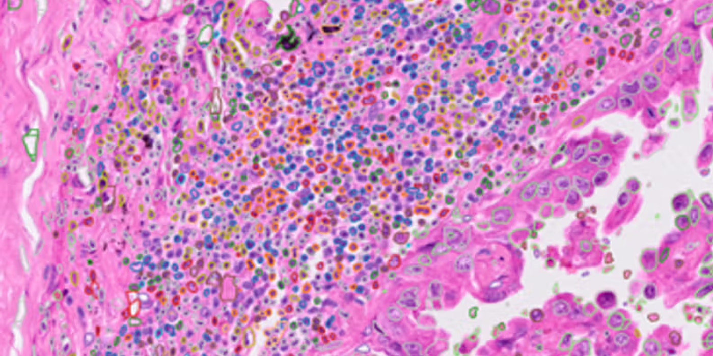
- Cell segmentation: Drawing boundaries around individual cells
- Cell classification: Identifying what type each cell is (tumor cell, immune cell, fibroblast, etc.)
The Impact: Democratizing Spatial Analysis
By working with standard H&E slides, CytoProfile makes advanced cellular analysis:
- Accessible: No need for expensive spatial profiling equipment
- Scalable: Can analyze thousands of archived slides
- Integrable: Fits into existing pathology workflows
- Retrospective: Works on historical tissue samples
This is particularly exciting for research institutions that may not have access to cutting-edge spatial biology platforms but have vast archives of H&E slides waiting to be analyzed.
What You Can Do With It
Once CytoProfile has analyzed an H&E slide, researchers can:
- Map cellular neighborhoods: See which cell types cluster together
- Quantify heterogeneity: Measure how varied the cellular composition is
- Identify spatial patterns: Detect immune-excluded vs. immune-inflamed tumors
- Compare across cohorts: Analyze cellular architecture in large patient studies
The Bigger Picture
CytoProfile represents a growing trend in computational pathology: using AI to extract maximum information from minimal input. Rather than requiring researchers to generate new, expensive data, it leverages the wealth of routine histology images already being created.
It's not replacing advanced techniques like multiplexing - those still provide the ground truth for training. Instead, it's extending their reach, making spatial cellular analysis practical for everyday research and potentially, one day, routine clinical use.
The Bottom Line: CytoProfile bridges the gap between cutting-edge spatial biology and routine pathology, bringing detailed cellular characterization to the H&E slides that are already the foundation of cancer diagnosis. It's about making advanced insights accessible, not just available.